On Monday, December 2, at Chicago’s McCormick Place Convention Center, the annual RSNA Conference included more sessions than ever that were focused on artificial intelligence (AI), and its application to radiological practice. Among numerous sessions today that involved AI was a session held at 3 PM local time, and entitled “Informatics (Artificial Intelligence: Triage, Screening, Quality).” While the title of that session didn’t fully convey its content, much was shared by its first two presenters that was enlightening for radiologists and other radiology-related professionals, around controlled studies assessing the effectiveness of AI in specific clinical practice situations.
The first two presenters in that session were S.S. Jayadeepa, M.D. and Melissa A. Davis, M.D. Both looked at AI in clinical practice.
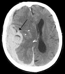
Dr. Jayadeepa, a practicing radiologist in Bangalore, India, shared the results of a study that she conducted in her local hospital, under the headline, “Automated AI Detection and Measurement of Midline Shift on Head CT Scans in an Emergency Teleradiogy Set-Up.” As the Wikipedia entry on midline shift explains, “Midline shift is a shift of the brain past its center line. The sign may be evident on neuroimaging such as CT scanning. The sign is considered ominous because it is commonly associated with a distortion of the brain stem that can cause serious dysfunction evidenced by abnormal posturing and failure of the pupils to constrict in response to light. Midline shift is often associated with high intracranial pressure (ICP), which can be deadly. In fact, midline shift is a measure of ICP; presence of the former is an indication of the latter. Presence of midline shift is an indication for neurosurgeons to take measures to monitor and control ICP. Immediate surgery may be indicated when there is a midline shift of over 5 mm. The sign can be caused by conditions including traumatic brain injury, stroke, hematoma, or birth deformity that leads to a raised intracranial pressure.”
As noted on her initial slide, the purpose of Dr. Jayadeepa’s study was “To evaluate the efficiency of an AI program using complex neural networks and deep learning algorithms for the detection and measurement of midline shift on non-contrast computed tomography examinations of the head.”
“In our teleradiology setup that reads over 300 PET CT head scans a day,” Jayadeepa told her audience, “it was very important to prioritize critical findings, such as intracranial hemorrhage, acute infarcts, mass effect, and midline shift.” As a result, she noted, “We looked at 163 non-contrast pre-operative non-contrast adult CT exams of the head. We found 93 cases positive for midline shift of more than 3 mm and 70 cases negative for midline shift of 3 mm.” She and her colleagues set the algorithm to flag an alert when a midline shift of more than 3 millimeters was detected. She and her colleagues found an accuracy of 95.15 percent.
The practical impact of this was significant. “The immediate detection and accurate measurement of midline shift on head CT exams is key to prompt patient triage and management in the emergency setting,” Jayadeepa explained. “An AI algorithm demonstrated promising results in both detection and quantification of midline shift, thereby allowing for prioritization of radiologist review, accelerated critical value communication, and enhanced patient care.”
Meanwhile, Dr. Davis, medical director, clinical redesign at Yale New Haven Health and chief of emergency radiology at the Yale University School of Medicine, presented on the topic, “Utilizing Machine Learning to Improve ED and In-Patient Throughput in cases of Acute Intracranial Hemorrhage by Non-Contrast Head CT.”
In that study, she and her colleagues looked at an important phenomenon, that of intracranial hemorrhage. As the Wikipedia entry on intracranial hemorrhage explains, “Intracranial hemorrhage (ICH), also known as intracranial bleed, is bleeding within the skull.[1] Subtypes are intracerebral bleeds (intraventricular bleeds and intraparenchymal bleeds), subarachnoid bleeds, epidural bleeds, and subdural bleeds. Intracerebral bleeding affects 2.5 per 10,000 people each year. Intracranial hemorrhage is a serious medical emergency because the buildup of blood within the skull can lead to increases in intracranial pressure, which can crush delicate brain tissue or limit its blood supply. Severe increases in intracranial pressure (ICP) can cause brain herniation, in which parts of the brain are squeezed past structures in the skull.”
“Intracranial hemorrhage is a high-incidence phenomenon,” Dr. Davis emphasized, “with 24.6 per 100,000 person-years; there are more than 40,000 cases of intracranial hemorrhage in the U.S. every year.” What’s more, she noted, “The 30-day mortality rate runs between 35 and 52 percent; and half of that mortality occurs within 24 hours.”
In this case, Davis noted, “We wanted to look at the impact of turnaround time because of the use of AI to trigger the prioritization of certain cases, compared to the standard ‘first-in-first-out’ approach to prioritization” of such cases. So, she said, “We implemented a machine learning algorithm based on a convolutional neural network that detects hyperdense abnormalities on head CT. The machine learning algorithm was incorporated across 29,000 cases. A prior study of the platform that provided the algorithm had found a sensitivity rate of 99 percent, a specificity rate of 95 percent, and an accuracy rate of 98 percent.
According to the terms of the study, “When it came to flagged cases”—cases in which the algorithm alerted the radiologist of the probability of an intercranial hemorrhage—radiologists “could choose to interact with the notification or do first-in-first-out. They could launch it in the PACS viewer or not, and dictate the case or not.”
Importantly, Davis reported, there was “a significant decrease in turnaround time, from 51.2 minutes, to 45.9 minutes, across the system, and from 53.0 minutes to 44.3 minutes, in our level 1 trauma center.” The leveraging of the algorithm also resulted in a significant decrease in length of stay.
“We hypothesized that patients were able to be triaged more expeditiously, especially in the ED,” Davis told her audience. “This may be especially helpful in EDs without radiologists or without routine immediate oversight of imaging, as in outpatient cases. And, she said, further research needs to be done to consider the effects on other acute and non-acute situations.
The decrease in turnaround time that resulted from the utilization of the machine learning-driven algorithm, Davis said, shows how useful AI and ML algorithms might be in a number of clinical situations going forward.


